Fun at the Orthopedist's
 Phil Pilgrim (PhiPi)
Posts: 23,514
Today I went to the orthopedist again to have my fractured foot x-rayed to see if there's been any progress at healing. They've got a really high-tech x-ray set-up: no film, everything is digital. Film packs have been replaced with an x-ray-sensitive tablet that works like film, except that the image is transmitted directly to a PC in the ante-room. One of the interesting facets of this arrangement is that multiple exposures can be taken without having to change film packets. This means that multiple images can be obtained from slightly different angles without disturbing the overall position of the subject on the tablet.
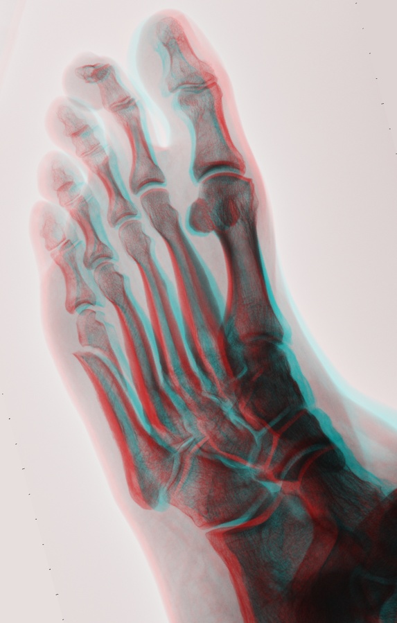
To me, this immediately suggested 3D. Take an image at two slightly different angles and use the two images to create a 3D anaglyph that can be viewed with red-green or red-blue glasses. In my case, three images have been taken each time I visit: a plan view, a profile view, and an oblique view. This time I convinced the x-ray tech to take two oblique-view images, between which I would lean my foot at a slightly different angle. She was totally into it and agreed readily. Here's the resulting anaglyph that I produced from the CDROM obtained from the clinic and which can be viewed with 3D glasses:

Although I'm sure that skilled radiologists do not need the extra dimension to analyze x-ray images, it gave me a new perspective on the overall shape of things. BTW, although the fracture still looks like a clean break, I was assured that there is new growth spanning the gap. It just takes time and, fortunately, I'm free to engage in most of the activities that I enjoy without restriction.
Who knew that a trip to the doc could have its geeky upside?
-Phil
P.S. If your 3D glasses have red on the left and blue on the right, you will see an image that looks like the top of a left foot, which is correct. Reversing those two filters will yield an image that looks like the bottom of a right foot.
To me, this immediately suggested 3D. Take an image at two slightly different angles and use the two images to create a 3D anaglyph that can be viewed with red-green or red-blue glasses. In my case, three images have been taken each time I visit: a plan view, a profile view, and an oblique view. This time I convinced the x-ray tech to take two oblique-view images, between which I would lean my foot at a slightly different angle. She was totally into it and agreed readily. Here's the resulting anaglyph that I produced from the CDROM obtained from the clinic and which can be viewed with 3D glasses:
Although I'm sure that skilled radiologists do not need the extra dimension to analyze x-ray images, it gave me a new perspective on the overall shape of things. BTW, although the fracture still looks like a clean break, I was assured that there is new growth spanning the gap. It just takes time and, fortunately, I'm free to engage in most of the activities that I enjoy without restriction.
Who knew that a trip to the doc could have its geeky upside?
-Phil
P.S. If your 3D glasses have red on the left and blue on the right, you will see an image that looks like the top of a left foot, which is correct. Reversing those two filters will yield an image that looks like the bottom of a right foot.


Comments
OUCH!
-Phil
Yes, a friend of mine said he could fix it by lashing the two halves together with dissolving suture material. 'Gave me my choice of whiskey for the anesthetic, too!
_______
Seriously, though, at this scale the growth that's beginning to span the gap does not show up, but it's there in the full-size image. It's just not very dense yet and won't achieve full fill and hard calcification for another 6 to 9 months. Meanwhile, there's no discomfort, and the only activities I have to avoid are impact sports, like running.
-Phil
Very nifty experiment! Way to use that adult onset A D D - I think my daughter has some red/blue 3d glasses someplace.
They might want to hang it on a wall together with some glasses.
Fun for the kids and all that...
This is novel, and over looked by the med community, since this is not already available at every x-ray station in the world.
Just printing the glasses for the doctor to give away at each session would make millions. (from revenues for ads printed on the glasses).
Thank me later when you are rich(er than you are now).
Ha! I'll bet your significant other stomped on your foot 'cause you spend too much at the Parallax store!
Heck, I even want one, you x-ray technician-sweet-talking devil.
I don't think Browser has enough mass to inflict such a wound
Jim
Looks like you really stubbed that second toe good. What's going on there?
PS
Is the red-blue orientation right? The standard is the red lens on the (observer's) left eye.
I used Corel PhotoPaint. The left and right images were converted to grayscale, then the right image was duplicated, leaving three images. These were then recombined as RGB channels of the same image (i.e. red and green+blue = cyan). After that step I rotated the result a bit to make sure the L/R offset was mostly horizontal, since I had misaligned the images a little vertically.
The second toe thing is called "hammer toe", an inherited trait, according to the orthopedist, and not due to injury or ill-fitting shoes as many believe.
-Phil
That would be a "mallet toe" my day job is a Podiatrist
I wish I had some 3D glasses to view that X-Ray, very cool idea except for the extra rads.
Thanks, BTW, for correcting my terminology.
-Phil